Home

Testicular Ultrasound: What You Need to Know About the Procedure and Its Benefits
In this Article

Testicular Ultrasound: What You Need to Know About the Procedure and Its Benefits
Updated on 8 June 2023
Imagine having the power to look inside your body, observing the hidden mysteries within. Well, with the incredible technology of testicular ultrasound, that power becomes a reality. Testicular ultrasound is a non-invasive imaging procedure that allows healthcare professionals to examine the testicles and surrounding structures with remarkable clarity.
This article will explain every aspect of testicular ultrasound, exploring its procedure, benefits, and the crucial information it can reveal. So, fasten your seatbelts as we embark on a journey of discovery through testicular ultrasound.
What is a Testicular Ultrasound?
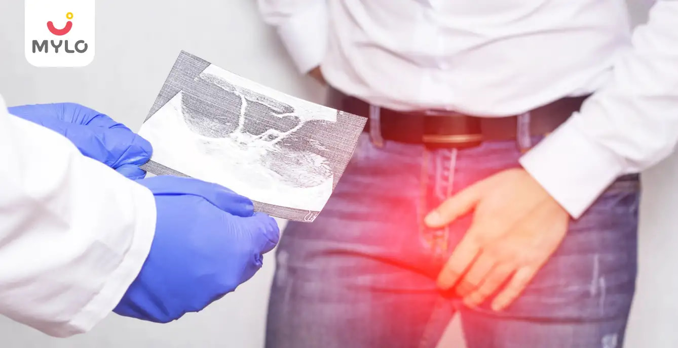
A testicular ultrasound is a painless imaging procedure that uses sound waves to create detailed images of the testicles and nearby structures. It involves using a small handheld device called a transducer, which emits high-frequency sound waves that bounce off the tissues and create echoes. These echoes are then converted into real-time images on a computer screen, allowing healthcare professionals to visualize the internal structures of the testicles.
Testicular doppler ultrasound is a valuable diagnostic tool that helps detect and evaluate various conditions affecting the testicles, such as tumors, infections, and abnormalities in blood flow. It provides valuable information to guide medical decisions and ensure optimal reproductive health in males.
You may like: Top 4 Kinds of Infertility Treatments to Cure Male Infertility
Who Needs a Testicular Ultrasound?
A testicular doppler ultrasound may be recommended for individuals experiencing symptoms or having certain risk factors that require further evaluation of the testicles. Some common reasons why someone may need a testicular ultrasound include:
1. Testicular pain or discomfort
If you're experiencing persistent pain, swelling, or lumps in the testicles, a testicular ultrasound can help identify the underlying cause, such as testicular torsion, epididymitis, or a testicular mass.
2. Testicular trauma
If you've had an injury or trauma to the testicles, a testicular ultrasound can assess for any structural damage, bleeding, or other complications.
3. Abnormal testicular findings
Suppose your healthcare provider detects an abnormality during a physical examination, such as an enlarged testicle or a suspicious lump. In that case, a testicular ultrasound can provide more detailed information about the nature of the abnormality.
4. Infertility concerns
Testicular ultrasound can evaluate the testicles for potential causes of male infertility, such as varicoceles (enlarged veins in the scrotum) or blockages in the sperm ducts.
5. Monitoring testicular conditions
Regular testicular ultrasounds may be recommended for individuals with known testicular conditions, such as testicular cancer or hydrocele, to monitor the condition and assess treatment effectiveness.
It's important to note that the decision to undergo a testicular ultrasound is made on a case-by-case basis by your healthcare provider, who will consider your symptoms, medical history, and other relevant factors.
You may like: Are You Aware of These Common Male Infertility Problems? What Are the Signs, Symptoms & Causes of It?
How is a Testicular Ultrasound Performed?
You will be asked to lie on your back on an examination table during a normal testicular doppler ultrasound. Here is a general overview of how the procedure is performed:
1. Preparation
You may be asked to remove your clothing from the waist down and wear a hospital gown. This allows easy access to the scrotum for the ultrasound.
2. Gel application
A clear gel will be applied to the scrotum. This gel helps the ultrasound probe make better contact with the skin and allows the sound waves to transmit more effectively.
3. Ultrasound probe
The healthcare provider gently moves a handheld ultrasound probe, called a transducer, over the scrotum. The transducer emits high-frequency sound waves that bounce off the tissues in the scrotum, creating images on a computer monitor.
4. Image capture
The healthcare provider will capture images of the testicles, epididymis, and surrounding structures. They may apply slight pressure with the transducer to obtain more explicit images or change the position of the scrotum if necessary.
5. Evaluation
The healthcare provider or a trained technician will evaluate the ultrasound images in real-time. They will look for any abnormalities, such as masses, cysts, or changes in blood flow.
6. Completion
Once the necessary images are obtained, the gel will be wiped off, and you can usually resume your normal activities immediately after the procedure.
The entire testicular ultrasound procedure is typically painless and takes 15 to 30 minutes to complete. You may feel slight pressure or coolness from the gel during the examination, but it should not be uncomfortable.
How to Prepare for a Testicular Doppler Ultrasound?
To prepare for a testicular Doppler ultrasound, you may need to follow specific instructions provided by your healthcare provider. Here are some general guidelines to help you prepare for the procedure:
1. Clothing
Wear comfortable and loose-fitting clothing on the day of the ultrasound. It is advisable to wear clothing that allows easy access to the scrotum area, as you may be asked to remove your pants or underwear during the examination.
2. Hygiene
It is essential to maintain proper hygiene before the procedure. Make sure to clean the genital area thoroughly to ensure accurate results and to avoid any discomfort during the examination.
3. Medications
Inform your healthcare provider about current medications, including over-the-counter drugs and supplements. They may provide specific instructions regarding the intake of medications before the test.
4. Avoid lotions or oils
Before the ultrasound, refrain from applying lotions, oils, or powders to the scrotum area. These substances can interfere with the ultrasound waves and affect the quality of the images.
5. Follow fasting instructions
Depending on the purpose of the routine testicular Doppler ultrasound, you may be asked to fast for a specific period before the procedure. This means avoiding food and drink, except water, for the instructed duration.
6. Questions and concerns
If you have any questions or concerns about the procedure, don't hesitate to discuss them with your healthcare provider beforehand. They can provide you with more specific instructions based on your case.
What are the Benefits of a Testicular Ultrasound?
A testicular Doppler ultrasound offers several benefits in diagnosing and evaluating conditions related to the testicles. Here are some key advantages of this procedure:
1. Non-invasive
Testicular ultrasound is a non-invasive imaging technique that does not involve any needles, incisions, or radiation exposure. It uses harmless sound waves to create detailed images of the testicles and surrounding structures.
2. Diagnostic tool
A testicular ultrasound helps diagnose conditions such as testicular torsion, testicular masses or tumors, epididymitis, hydrocele, varicocele, and other abnormalities. It allows healthcare professionals to visualize the testicles and assess their size, shape, blood flow, and any presence of masses or abnormalities.
3. Guiding treatment decisions
The detailed images obtained from a testicular ultrasound aid in guiding treatment decisions. It helps healthcare providers determine the most appropriate action, whether monitoring a condition, planning surgery, or recommending further diagnostic tests.
4. Assessing fertility concerns
Testicular ultrasound can assess the health and functionality of the testicles, which is crucial for evaluating male fertility. It can identify issues such as varicoceles (enlarged veins in the scrotum), which may affect sperm production and quality.
5. Monitoring response to treatment
For individuals undergoing treatment for testicular conditions, such as tumors or infections, testicular ultrasound can be used to monitor the effectiveness of the treatment and track any changes or improvements over time.
6. Quick and convenient
Testicular ultrasound is typically a quick and convenient procedure that can be performed in a medical facility or imaging center. It is generally well-tolerated and does not require any recovery time.
Conclusion
In conclusion, testicular ultrasound is a valuable diagnostic tool that offers numerous benefits in assessing and evaluating testicular health. With its ability to identify abnormalities, guide treatment decisions, and provide valuable insights into testicular health, testicular doppler ultrasound plays a crucial role in maintaining reproductive well-being and addressing potential concerns. If you have any testicular-related symptoms or concerns, consulting with a healthcare professional and considering a testicular ultrasound can provide valuable information and peace of mind.
References
1. Kühn, A. L., Scortegagna, E., Nowitzki, K. M., & Kim, Y. H. (2016). Ultrasonography of the scrotum in adults. Ultrasonography, 35(3), 180–197.
2. Yusuf, G. T., & Sidhu, P. S. (2013). A review of ultrasound imaging in scrotal emergencies. Journal of Ultrasound, 16(4), 171–178.


Written by
Madhavi Gupta
Dr. Madhavi Gupta is an accomplished Ayurvedic doctor specializing in Medical content writing with an experience of over 10 years.
Read MoreGet baby's diet chart, and growth tips

Related Articles
Related Questions
Influenza and boostrix injection kisiko laga hai kya 8 month pregnancy me and q lagta hai ye plz reply me

Hai.... My last period was in feb 24. I tested in 40 th day morning 3:30 .. That is faint line .. I conculed mylo thz app also.... And I asked tha dr wait for 3 to 5 days ... Im also waiting ... Then I test today 4:15 test is sooooo faint ... And I feel in ma body no pregnancy symptoms. What can I do .

Baby kicks KB Marta hai Plz tell mi

PCOD kya hota hai

How to detect pcos

RECENTLY PUBLISHED ARTICLES
our most recent articles

Symptoms of Low AMH to Watch Out For: A Health Alert for Women Trying to Conceive

Medical Procedures
Hysteroscopy: Everything You Need to Know About This Minimally Invasive Procedure

Ayurveda & Homeopathy
Dalchini: How This Herb Can Make Way From Your Spice Rack to Your Medicine Cabinet

Health & Wellness
Fenugreek Powder: Health Benefits of Fenugreek From Your Kitchen to Your Medicine Cabinet

Lifestyle
Moringa Powder: The Superfood You Need in Your Diet for a Healthy Lifestyle

Illnesses & Infections
Genital Herpes: Causes, Symptoms, Risks & Treatment
- Ashokarishta: All You Need to Know About This Miracle Tonic for Women
- 10 Amazon Prime Movies to Look Forward to in 2023
- 10 Best Netflix Movies to Watch Out For in 2023
- Fertility Yoga: A Natural Solution to Boost Your Chances of Conception
- How to Get Regular Periods Naturally: Ayurvedic Herbs, Lifestyle Changes & Homeopathy
- Lodhra: The Wonder Herb for Women's Health
- Malabsorption Syndrome: Types, Causes, Symptoms, & Treatment
- Top 10 Short Bedtime Stories for Kids
- RH Incompatibility in Pregnancy - Causes, Symptoms & Treatments
- Patent Ductus Arteriosus (PDA) Symptoms & Treatment
- Why Babies Cry After Birth?
- IVF Process Step by Step Timeline: What to Expect During Your Fertility Journey
- Tracheoesophageal Fistula: Causes, Symptoms, Risks & Treatment
- Stillbirth: Cause, Symptoms, Risks & Prevention


AWARDS AND RECOGNITION

Mylo wins Forbes D2C Disruptor award

Mylo wins The Economic Times Promising Brands 2022
AS SEEN IN


- Mylo Care: Effective and science-backed personal care and wellness solutions for a joyful you.
- Mylo Baby: Science-backed, gentle and effective personal care & hygiene range for your little one.
- Mylo Community: Trusted and empathetic community of 10mn+ parents and experts.
Product Categories
baby carrier | baby soap | baby wipes | stretch marks cream | baby cream | baby shampoo | baby massage oil | baby hair oil | stretch marks oil | baby body wash | baby powder | baby lotion | diaper rash cream | newborn diapers | teether | baby kajal | baby diapers | cloth diapers |


