Home

Scans & Tests

Fetal Echo Test in Pregnancy: A Diagnostic Tool for Detecting Heart Defects in the Womb
In this Article

Scans & Tests
Fetal Echo Test in Pregnancy: A Diagnostic Tool for Detecting Heart Defects in the Womb
Updated on 25 April 2023
Fetal echocardiography, also known as fetal echo, is a specialized ultrasound technique used to evaluate the heart of a developing fetus. This diagnostic tool has revolutionized prenatal care and has become an essential part of the routine prenatal screening process. In this article, we will understand in detail about fetal echo test in pregnancy, why it is important, who needs it, and how it is performed.
What is a Fetal Echo Scan?
A fetal echo test is a specialized ultrasound that is performed to evaluate the structure and function of the fetal heart. The scan provides detailed images of the heart, including the chambers, valves, and blood vessels. It also measures the blood flow through the heart, which can help detect any anomalies.
It is a non-invasive, painless diagnostic test that uses high-frequency sound waves to create images of the developing heart of a fetus. This technique is like a regular ultrasound, but it focuses specifically on the heart. The earliest time a fetal echo scan can be performed is at around 18-20 weeks of gestation when the fetal heart is fully developed and can be visualized on an ultrasound. However, it can be performed at any time during the pregnancy if the need arises.
You may also like: How to Check Baby's Heartbeat During Pregnancy at Home
Why is Fetal Echocardiography Important?
Fetal echocardiography is a crucial tool in the diagnosis of congenital heart defects, which are the most common birth defects affecting approximately 1 in every 100 births. Early detection of heart defects can help in the management of pregnancy and can also prepare parents for the possibility of a child with a heart condition.
In some cases, early detection can also provide an opportunity for in-utero treatment or intervention. Fetal echo can also be used to monitor fetuses that are at risk for heart problems due to genetic or other factors.
You may also like: How Foul Air May Affect Fetal Heart Development
Who Needs a Fetal Echo Test in Pregnancy?
A fetal echo scan is recommended for pregnant women who have a higher risk of having a baby with a heart defect. This includes women who have a family history of congenital heart defects, have previously had a child with a heart defect, have certain medical conditions such as diabetes, lupus, or phenylketonuria, or have taken certain medications during pregnancy.
In addition, fetal echocardiography may be recommended for women who have abnormal findings on routine prenatal ultrasound or have other risk factors identified during their pregnancy.
How is Fetal Echocardiography Performed?
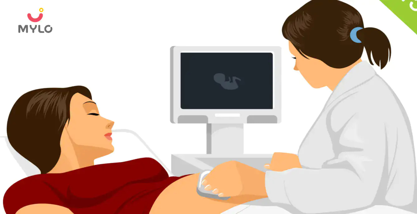
Fetal echo is a painless and non-invasive procedure that does not require any preparation on the part of the mother. The mother is asked to lie down on a table, and a gel is applied to her abdomen to help the transducer glide smoothly over the skin. The transducer is moved over the mother's abdomen to obtain images of the fetal heart. The procedure usually takes around 30-45 minutes.
What Can be Detected Through Fetal Echo?
Fetal echo test in pregnancy can detect a wide range of heart defects, including structural abnormalities, functional abnormalities, and abnormalities in blood flow. Some of the most common heart defects that can be detected through fetal echo include:
-
atrial and ventricular septal defects
-
coarctation of the aorta
-
tetralogy of Fallot
-
transposition of the great arteries
-
abnormalities in the heart rhythm and function
You may also like: Why is the Second Trimester Anomaly Scan Important During Pregnancy?
Interpretation of Fetal Echocardiography Results
The results of a fetal echo scan are interpreted by a trained specialist who is experienced in fetal heart evaluation. The results are usually reported as normal or abnormal, and if there are any abnormalities, further testing may be recommended. In some cases, the specialist may also recommend a follow-up scan to monitor the development of the fetal heart. If a heart defect is detected, the specialist will discuss the management and treatment options with the parents.
Risks and Limitations of Fetal Echo Test
Fetal echo scan is a safe and non-invasive procedure that does not pose any risks to the mother or the fetus. However, like all diagnostic tests, it does have some limitations. Fetal echo test in pregnancy can only detect heart defects that are present at the time of the scan, and it cannot predict any heart problems that may develop later in the pregnancy or after birth. In addition, fetal echocardiography is highly dependent on the skill and experience of the technician performing the scan, and there is a risk of false-positive or false-negative results.
You may also like: What Are the Common Tests You Will Have During Your Pregnancy?
Conclusion
In conclusion, fetal echocardiography is a safe and non-invasive procedure that can detect a wide range of heart defects in the developing fetus. If you are pregnant and have any concerns about your baby's heart health, talk to your healthcare provider about the possibility of a fetal echo. Early detection and management of congenital heart defects can make a significant difference in the health and well-being of your child.


Written by
Ravish Goyal
Official account of Mylo Editor
Read MoreGet baby's diet chart, and growth tips

Related Articles
Related Topics
RECENTLY PUBLISHED ARTICLES
our most recent articles

Symptoms & Illnesses
Bedwetting (Nocturnal Enuresis): Causes, Symptoms & Treatment

Skin Changes
Birthmark: Types, Causes, Risks & Treatment

Emotions & Behaviour
Behaviour Therapy: Benefits, Types & Techniques

Feeding Schedule
How Long Does Breast Milk Last at Room Temperature?
Home Remedies
Thrush: Causes, Symptoms, Treatment, and More

Illnesses & Infections
Childhood Asthma: Symptoms, Causes & Treatment
- Reflux in Baby: Symptoms, Causes & Treatment
- Pre Eclampsia: Meaning, Causes & Symptoms
- Baby Diarrhea: Causes, Symptoms & Treatment
- Bronchiolitis: Causes, Symptoms & Treatment
- Pelvic Pain in Pregnancy: Symptoms & Treatment
- Saliva During Pregnancy: Causes & Prevention
- Effective Ways to Treat Jaundice in Children: Expert Tips for a Speedy Recovery
- 10 Best Original Movies to Watch on Netflix
- Flu, Change of Season or New Covid Variant, XBB.1.16- What’s Causing These Symptoms?
- 5 Ways In Which Music Can Boost Your Baby's Brain Development
- How to Stop Baby Hiccups: Everything You Need to Know
- “Staying Active and Healthy: The Benefits of Safe Exercise During Pregnancy”
- Appendicitis In Pregnancy Symptoms, Diagnosis & Surgery
- 5 Common Myths Busted About Baby Sleep


AWARDS AND RECOGNITION

Mylo wins Forbes D2C Disruptor award

Mylo wins The Economic Times Promising Brands 2022
AS SEEN IN

- Mylo Care: Effective and science-backed personal care and wellness solutions for a joyful you.
- Mylo Baby: Science-backed, gentle and effective personal care & hygiene range for your little one.
- Mylo Community: Trusted and empathetic community of 10mn+ parents and experts.
Product Categories
baby carrier | baby soap | baby wipes | stretch marks cream | baby cream | baby shampoo | baby massage oil | baby hair oil | stretch marks oil | baby body wash | baby powder | baby lotion | diaper rash cream | newborn diapers | teether | baby kajal | baby diapers | cloth diapers |


